Embark on a captivating journey into the realm of anatomy with our “Inferior View of Skull Quiz.” This interactive experience will delve into the intricate structures that form the base of the skull, providing a comprehensive understanding of its significance in medical examinations, comparative anatomy, and developmental biology.
Prepare to unravel the mysteries of the occipital bone, temporal bones, and sphenoid bone, as we explore the foramina and fissures that adorn the inferior view of the skull. Discover the functions and relationships of the foramen magnum, condylar fossae, and jugular foramina, gaining insights into their clinical relevance and developmental processes.
Skull Anatomy Overview: Inferior View Of Skull Quiz
The skull, also known as the cranium, is a complex structure that encloses and protects the brain and other delicate organs of the head. It consists of 22 bones that are joined together by sutures, which are immovable joints. The skull can be divided into two main parts: the cranium and the facial skeleton.
The cranium is the upper part of the skull and it houses the brain. The facial skeleton is the lower part of the skull and it supports the facial structures.
The inferior view of the skull quiz is a great way to test your knowledge of the anatomy of the skull. The quiz covers a variety of topics, including the bones of the skull, the foramina of the skull, and the muscles of the skull.
You can learn more about the skull by reading nri meaning oil and gas . The inferior view of the skull quiz is a great way to test your knowledge of the anatomy of the skull.
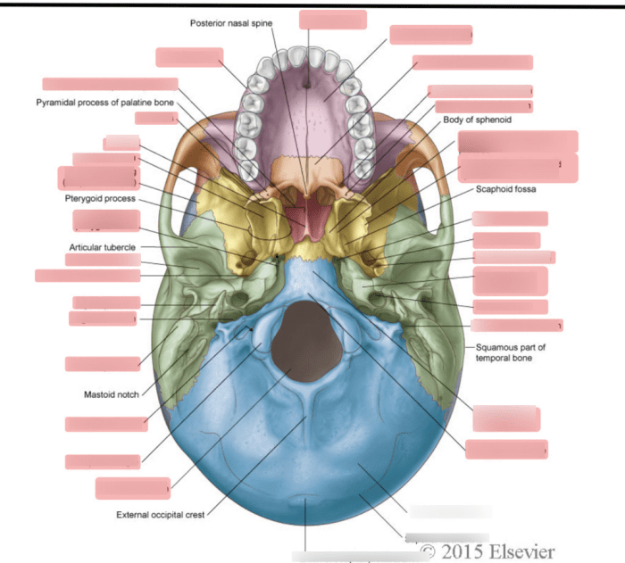
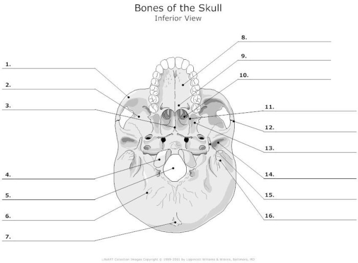
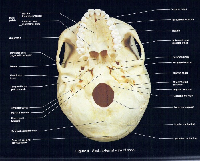
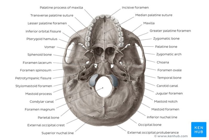
The inferior view of the skull reveals several important structures. These include the foramen magnum, which is a large opening at the base of the skull through which the spinal cord passes. The occipital condyles, which are two projections on either side of the foramen magnum, articulate with the first vertebra of the spinal column.
The styloid processes, which are two slender projections located on either side of the foramen magnum, provide attachment points for muscles.
The inferior view of the skull also provides a good view of the paranasal sinuses. These are air-filled cavities located within the facial bones. The paranasal sinuses help to lighten the skull and they also produce mucus, which helps to keep the nasal passages moist.
Diagram of the Skull
[Provide a diagram or illustration of the skull, highlighting the inferior view.]
Inferior View of the Skull
The inferior view of the skull provides a detailed look at the structures that form the base of the cranium. These structures play crucial roles in supporting the brain, protecting vital organs, and facilitating various functions related to the head and neck.
Bones of the Inferior View
The inferior view of the skull is primarily formed by three bones:
- Occipital bone:Located at the posterior aspect, it forms the back of the skull and provides attachment points for muscles and ligaments.
- Temporal bones:Situated on either side of the occipital bone, they contribute to the lateral and inferior aspects of the skull, housing the middle and inner ear structures.
- Sphenoid bone:Located anteriorly, it forms the central portion of the skull base and articulates with several other bones, contributing to the formation of the cranial cavity.
Foramina and Fissures
The inferior view of the skull also features several foramina and fissures, which allow for the passage of nerves, blood vessels, and other structures:
- Foramen magnum:A large opening in the occipital bone that allows the spinal cord to pass through and connect to the brain.
- Jugular foramen:Located on either side of the occipital bone, it transmits the jugular vein, cranial nerves, and carotid arteries.
- Hypoglossal canal:A small foramen on the occipital bone that transmits the hypoglossal nerve, responsible for tongue movement.
- Carotid canal:Found in the temporal bone, it transmits the internal carotid artery, supplying blood to the brain.
- Petrotympanic fissure:A narrow fissure in the temporal bone that connects the middle ear to the cranial cavity.
Structures on the Inferior View

The inferior view of the skull presents a range of anatomical structures crucial for the skull’s function and connection with the spine and other cranial elements. This view offers insights into the skull’s base, providing a comprehensive understanding of its structural components.
Foramen Magnum
The foramen magnum is a large, oval-shaped opening at the base of the skull. It serves as a passageway for the spinal cord to connect with the brainstem, allowing for the transmission of neural signals between the brain and the rest of the body.
The foramen magnum is surrounded by the occipital condyles, which articulate with the first cervical vertebra (atlas) to facilitate head movements.
Condylar Fossae
The condylar fossae are paired depressions located on either side of the foramen magnum. They accommodate the occipital condyles, forming synovial joints that allow for a wide range of head movements, including flexion, extension, and rotation. The condylar fossae are lined with cartilage to reduce friction during movement.
Jugular Foramina
The jugular foramina are paired openings located on either side of the foramen magnum. They transmit the jugular veins, which drain blood from the brain and head region, and the cranial nerves IX (glossopharyngeal), X (vagus), and XI (accessory). The jugular foramina are also associated with the carotid arteries, which supply blood to the brain.
Clinical Significance

The inferior view of the skull plays a crucial role in medical examinations and procedures due to its accessibility and the presence of various anatomical structures that can provide valuable clinical information.Abnormalities or injuries to the inferior view of the skull can manifest as palpable depressions, swelling, or discoloration, indicating potential fractures or underlying damage.
These abnormalities can be detected through physical examinations, imaging techniques like X-rays or CT scans, and endoscopic procedures.
Examination and Assessment
During a physical examination, the inferior view of the skull is palpated to assess for any irregularities, such as depressions or swellings, which may suggest a fracture or other injury. X-rays provide a two-dimensional view of the skull, allowing visualization of fractures, foreign bodies, or abnormalities in bone density.
CT scans offer more detailed cross-sectional images, enabling a thorough evaluation of the skull base and surrounding structures. Endoscopic procedures, such as nasal endoscopy or transoral robotic surgery, allow direct visualization of the skull base and can aid in diagnosing and treating conditions affecting this region.
Comparative Anatomy

The inferior view of the human skull exhibits distinct characteristics that distinguish it from other species, including mammals, reptiles, and birds. Understanding these variations provides insights into evolutionary adaptations and functional differences among different animal groups.
Mammals
- Mammals generally possess a broad and flattened inferior surface of the skull, with a prominent occipital condyle for articulation with the first cervical vertebra.
- The foramina and canals on the inferior view are similar to humans, including the jugular foramen, carotid canal, and hypoglossal canal.
- However, the size and shape of these structures can vary depending on the species and its specific adaptations.
Reptiles, Inferior view of skull quiz
- Reptiles have a narrower and more elongated inferior view of the skull compared to mammals.
- The occipital condyle is typically divided into two distinct condyles, which articulate with the atlas and axis vertebrae.
- The foramina and canals on the inferior view are often reduced in size and complexity compared to mammals, reflecting the differences in their sensory and nervous systems.
Birds
- Birds have a highly modified inferior view of the skull, with a reduced and fused structure known as the basitemporal plate.
- The occipital condyle is located more anteriorly and is often smaller in size compared to mammals and reptiles.
- The foramina and canals on the inferior view are limited and adapted to the unique sensory and feeding adaptations of birds.
Developmental Anatomy

The inferior view of the skull, also known as the basal view, develops through a complex series of ossification and fusion processes that begin in the embryonic stage.The initial stage involves the formation of the chondrocranium, a cartilaginous precursor to the skull, which lays the foundation for the future bony structures.
As the embryo develops, specific regions of the chondrocranium undergo ossification, gradually replacing the cartilage with bone.
Stages of Ossification and Fusion
*
-*Primary Ossification
The first bones to ossify are the occipital bone, temporal bones, and sphenoid bone. These bones form the base of the skull and provide attachment points for muscles and ligaments.
-
-*Secondary Ossification
Later in development, additional bones ossify within the cartilaginous framework. These include the palatine bones, maxillae, and nasal bones, which contribute to the formation of the hard palate and nasal cavity.
-*Fusion
As ossification progresses, the individual bones begin to fuse together. The sutures, or immovable joints, between the bones gradually disappear, creating a solid and continuous structure. This fusion process ensures the strength and stability of the skull.
FAQ Corner
What are the main bones that form the inferior view of the skull?
The occipital bone, temporal bones, and sphenoid bone.
What is the significance of the foramen magnum?
It allows the passage of the spinal cord from the skull to the vertebral column.
How does the inferior view of the skull differ between humans and other species?
There are variations in bone structure, foramina, and overall morphology, reflecting adaptations to different environments and lifestyles.