Any of 12 spinal bones crossword clue – In the intricate tapestry of the human body, the spinal column stands as a pivotal structure, housing and protecting the delicate spinal cord. Comprising 12 distinct vertebrae, this remarkable framework provides essential support, flexibility, and protection for our neural pathways.
Embark on a journey to unravel the enigma of the spinal column, delving into the anatomy, functions, and significance of each vertebra.
From the cervical vertebrae that cradle our heads to the coccygeal vertebrae that anchor our pelvic floor, each vertebra plays a unique role in the overall symphony of our musculoskeletal system. Join us as we explore the fascinating world of spinal bones, uncovering their intricate connections and vital contributions to our well-being.
Vertebrae
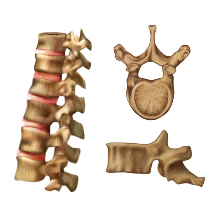
Vertebrae are the individual bones that make up the spinal column, also known as the backbone. They provide structural support for the body, protect the spinal cord, and allow for movement and flexibility.
Each vertebra has a complex anatomical structure, consisting of a body, pedicles, laminae, and various processes. The body is the main weight-bearing component and forms the front part of the vertebra. Pedicles are short, thick projections that extend from the back of the body and connect to the laminae.
Types of Vertebrae
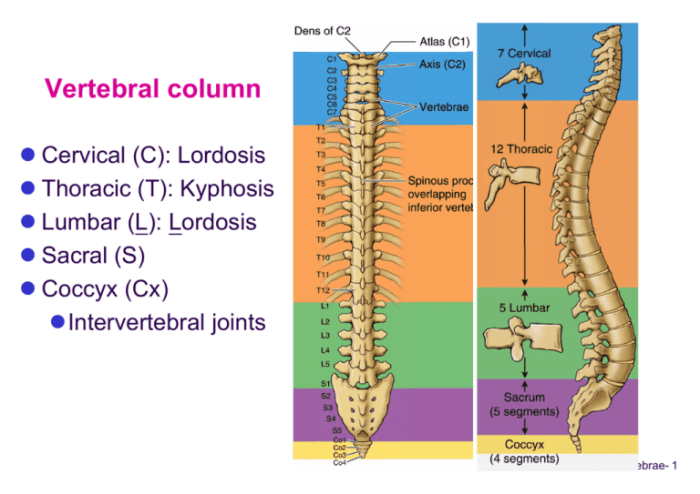
There are five types of vertebrae, each with distinct characteristics and locations within the spinal column:
- Cervical vertebrae (7):Found in the neck region, these vertebrae are the smallest and lightest, allowing for a wide range of head movements.
- Thoracic vertebrae (12):Located in the chest area, these vertebrae are larger and have facets for articulation with ribs.
- Lumbar vertebrae (5):Found in the lower back, these vertebrae are the largest and strongest, supporting the weight of the upper body.
- Sacral vertebrae (5):Fused together to form the sacrum, these vertebrae provide stability to the pelvis.
- Coccygeal vertebrae (4):Fused together to form the coccyx, these vertebrae are small and vestigial.
Spinal Column
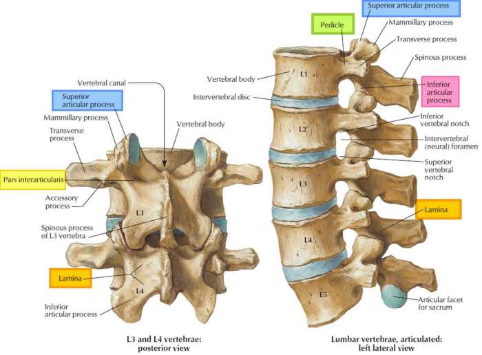
The spinal column, also known as the vertebral column, is a complex and vital structure that forms the central axis of the skeletal system. It is composed of a series of individual bones called vertebrae, which are stacked one upon another to form a flexible and supportive structure.
The spinal column serves several essential functions. It provides structural support for the body, allowing us to stand, walk, and move with ease. It also protects the delicate spinal cord, which runs through the center of the column and carries messages between the brain and the rest of the body.
Additionally, the spinal column provides attachment points for muscles and ligaments, which help to stabilize and control movement.
Vertebrae
The vertebrae are the individual bones that make up the spinal column. Each vertebra has a unique shape and structure, depending on its location in the column. The vertebrae are connected to each other by a series of ligaments and joints, which allow for flexibility and movement.
The spaces between the vertebrae are filled with intervertebral discs, which act as cushions to absorb shock and prevent the vertebrae from rubbing against each other.
Protection of the Spinal Cord
One of the most important functions of the spinal column is to protect the spinal cord. The spinal cord is a delicate bundle of nerves that runs from the brain down the center of the spinal column. It is responsible for transmitting messages between the brain and the rest of the body.
The spinal column provides a strong and protective casing around the spinal cord, shielding it from injury.
Support and Stability
The spinal column also provides support and stability for the body. It acts as a central axis around which the body can move and balance. The vertebrae are connected to each other by a series of ligaments and muscles, which help to stabilize the column and prevent it from buckling or collapsing.
Cervical Vertebrae
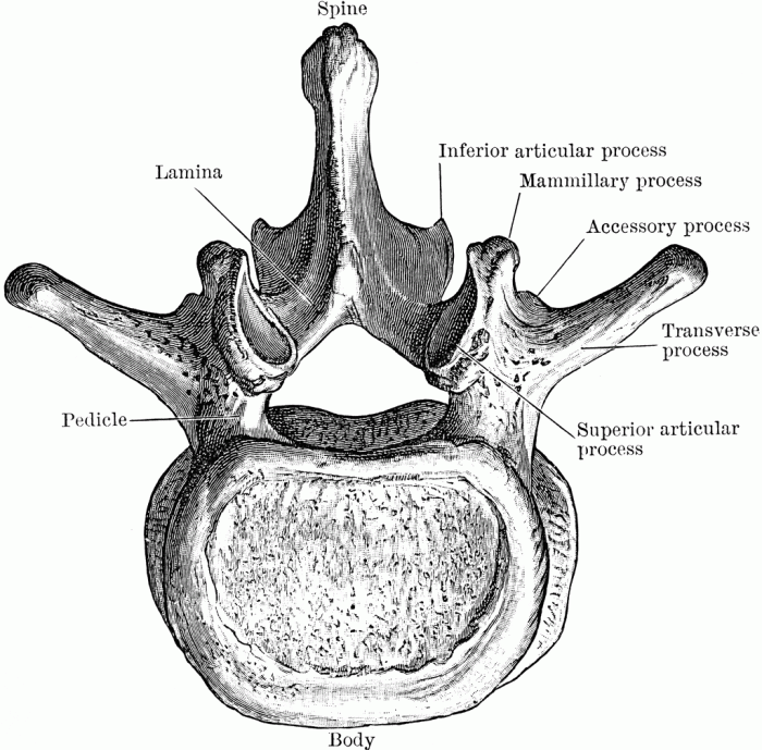
The cervical vertebrae, also known as the neck bones, are the seven vertebrae located in the neck region of the spinal column. They play a crucial role in supporting the head, facilitating neck movement, and protecting the spinal cord.
Each cervical vertebra has unique characteristics that contribute to its overall function. The first two cervical vertebrae, the atlas and axis, are highly specialized and allow for a wide range of head movements.
Atlas (C1)
- First cervical vertebra, located at the base of the skull
- Ring-shaped, with no body or spinous process
- Articulates with the occipital condyles of the skull, allowing for nodding (flexion/extension) and side-to-side (lateral flexion) head movements
Axis (C2)
- Second cervical vertebra, located above the atlas
- Has a prominent odontoid process (dens) that projects upward into the atlas
- Allows for rotation (pivoting) of the head
Remaining Cervical Vertebrae (C3-C7)
- Have a typical vertebral structure, with a body, pedicles, laminae, and spinous process
- Increase in size from C3 to C7
- Articulate with each other through facet joints, allowing for flexion, extension, lateral flexion, and some rotation
| Vertebra | Unique Characteristics | Role |
|---|---|---|
| Atlas (C1) | Ring-shaped, no body or spinous process | Nodding, side-to-side head movements |
| Axis (C2) | Odontoid process | Head rotation |
| C3-C7 | Typical vertebral structure | Flexion, extension, lateral flexion, some rotation |
Thoracic Vertebrae
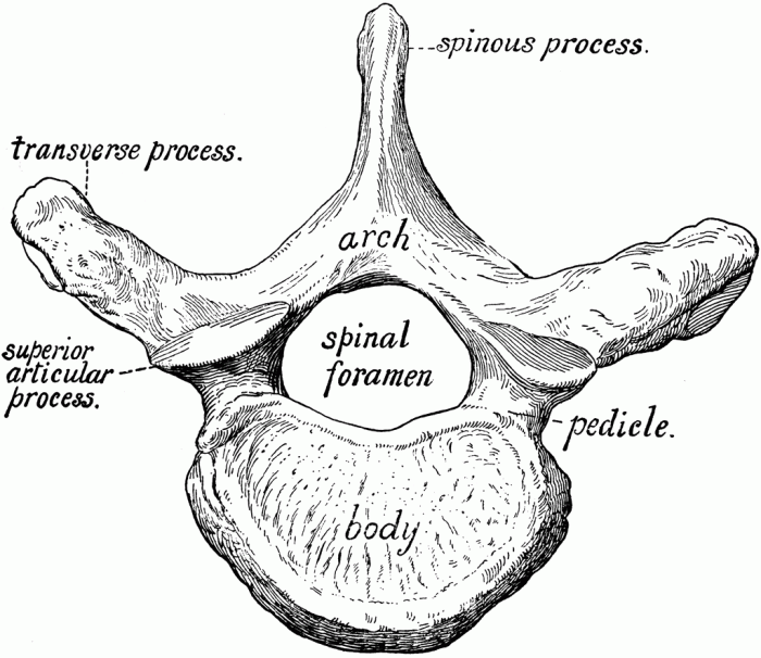
The thoracic vertebrae, comprising 12 bones, constitute the middle section of the spinal column and play a pivotal role in safeguarding the vital organs within the thoracic cavity. Each thoracic vertebra exhibits distinctive features that contribute to its overall functionality.
The thoracic vertebrae are responsible for providing structural support and protection to the heart and lungs, ensuring their optimal functioning. They facilitate the attachment of ribs, forming the thoracic cage, which serves as a protective enclosure for these delicate organs.
Unique Characteristics of Each Thoracic Vertebra
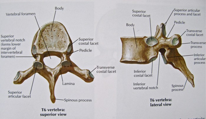
- Body:The vertebral body is heart-shaped and larger than those of the cervical vertebrae, reflecting the increased weight-bearing capacity required in this region.
- Pedicles:The pedicles are short and thick, providing stability and strength to the vertebral column.
- Laminae:The laminae are broad and flat, forming the roof of the vertebral foramen.
- Transverse Processes:The transverse processes are long and extend laterally, serving as attachment points for the ribs.
- Spinous Process:The spinous process is long and triangular, pointing inferiorly, providing attachment for muscles and ligaments.
- Facet Joints:The facet joints are located on the superior and inferior articular processes, allowing for movement between adjacent vertebrae.
Lumbar Vertebrae
The lumbar vertebrae, consisting of five bones numbered L1 to L5, are the largest and strongest vertebrae in the spinal column. They are located in the lower back and play a crucial role in supporting the weight of the upper body and facilitating movement.The
lumbar vertebrae are characterized by their robust bodies, which are thicker and wider than those of other spinal vertebrae. They also have shorter and broader spinous processes that project backward from the vertebral arches. The transverse processes of the lumbar vertebrae are long and extend laterally, providing attachment points for muscles that support the back and abdomen.The
fifth lumbar vertebra (L5) is the largest and strongest of the lumbar vertebrae. It has a prominent body and a thick, triangular spinous process. The L5 vertebra also has a large, flat articular surface on its inferior aspect, which articulates with the sacrum.The
lumbar vertebrae are essential for supporting the weight of the upper body and allowing for movement. They provide stability to the lower back and facilitate bending, twisting, and lifting. The lumbar vertebrae also protect the spinal cord and nerve roots that pass through the spinal canal.
- L1: The first lumbar vertebra is the smallest and most transitional of the lumbar vertebrae. It has features similar to both thoracic and lumbar vertebrae.
- L2: The second lumbar vertebra is larger than L1 and has a more pronounced lumbar character. It has a thicker body and shorter spinous process.
- L3: The third lumbar vertebra is the largest of the lumbar vertebrae. It has a robust body and a thick, triangular spinous process.
- L4: The fourth lumbar vertebra is similar in size to L3. It has a slightly smaller body and a shorter spinous process.
- L5: The fifth lumbar vertebra is the largest and strongest of the lumbar vertebrae. It has a prominent body and a thick, triangular spinous process.
The following diagram compares the size and shape of the lumbar vertebrae to other spinal vertebrae:[Image of a diagram comparing the size and shape of the lumbar vertebrae to other spinal vertebrae]
Sacral Vertebrae: Any Of 12 Spinal Bones Crossword Clue
The sacral vertebrae are five fused vertebrae located at the base of the spine. They form the posterior wall of the pelvis and provide stability to the spine and pelvis.
Fused Structure
The sacral vertebrae are fused together to form a triangular-shaped bone called the sacrum. The sacrum articulates with the ilium bones of the pelvis to form the sacroiliac joints.
Role in Pelvis Formation and Stability, Any of 12 spinal bones crossword clue
The sacral vertebrae play a crucial role in forming the pelvis and providing stability to the spine and pelvis. The sacrum forms the posterior wall of the pelvis, providing support for the pelvic organs. The sacroiliac joints allow for some movement between the sacrum and the ilium, providing stability to the pelvis and spine.
Differences between Sacral Vertebrae and Other Spinal Vertebrae
The sacral vertebrae differ from other spinal vertebrae in several ways:
| Characteristic | Sacral Vertebrae | Other Spinal Vertebrae |
|---|---|---|
| Number | 5 | 7 cervical, 12 thoracic, 5 lumbar |
| Fusion | Fused together to form the sacrum | Not fused |
| Shape | Triangular | Variable depending on region |
| Function | Form the posterior wall of the pelvis and provide stability | Support the spine and protect the spinal cord |
Coccygeal Vertebrae
The coccygeal vertebrae, also known as the tailbone, are the four smallest and most inferior vertebrae of the spinal column. They are located at the bottom of the sacrum and are rudimentary in structure, meaning they are underdeveloped and have lost their original function.The
coccygeal vertebrae play a role in providing support to the pelvic floor, which is a group of muscles and ligaments that supports the pelvic organs. The coccyx also serves as an attachment point for several muscles, including the gluteus maximus and the levator ani.
Diagram of Coccygeal Vertebrae
[Image of coccygeal vertebrae, labeled with their names and sizes.]
FAQ Resource
What is the function of the spinal column?
The spinal column serves as a protective sheath for the spinal cord, provides structural support for the body, and facilitates movement.
How many vertebrae are there in the spinal column?
There are a total of 33 vertebrae in the human spinal column, divided into five regions: cervical (7), thoracic (12), lumbar (5), sacral (5 fused), and coccygeal (4 fused).
What is the difference between a vertebra and a spinal bone?
Vertebrae are the individual bones that make up the spinal column, while a spinal bone refers to the entire structure formed by the interconnected vertebrae.